Author: [AUTHOR] Published on 10/1/2026 12:00:00 AMFROM THE ASCPT ONCOLOGY COMMUNITY
ctDNA Explained
By Andrew B. SyBing, Associate Director of Clinical Pharmacology & Pharmacometrics @ Pfizer Oncology
Note: AI was utilized to assist with the creation of the initial draft of this resource. The Oncology (ONC) Community Communications Team used the draft to create and format the final version that appears here.
A blood sample may not seem like an obvious place to look for information about a tumor. Yet, tumors can leave molecular traces in the bloodstream, including small fragments of their DNA. Those fragments, known as circulating tumor DNA, or ctDNA, are an emerging method to study cancer without relying exclusively on tissue biopsies.
ctDNA is currently being used to identify actionable genomic alterations and characterize resistance mechanisms. The promise of ctDNA applications is wide, including 1) monitoring treatment response, 2) detecting molecular residual disease, 3) assessing recurrence risk, and 4) providing early evidence of therapeutic activity. Because blood collection is convenient, the collection of ctDNA allows for observation of tumor biology as it changes over the course of treatment rather than relying on infrequent, labor-intensive tumor biopsies.
IN THIS ARTICLE
Most cells release small fragments of DNA as part of normal cell turnover. DNA fragments found outside cells in blood and other body fluids are collectively referred to as
cell-free DNA, or
cfDNA.
In patients with cancer, a portion of the cfDNA may originate from tumor cells. This tumor-derived fraction is called ctDNA. ctDNA can contain many of the molecular characteristics found within a tumor, including point mutations, copy-number changes, rearrangements, and other genomic alterations. However, ctDNA generally represents only a fraction of the total cfDNA circulating in plasma, and the amount present can vary substantially among patients and disease settings.
Analyzing ctDNA from a blood sample is one form of liquid biopsy. Unlike a conventional tissue biopsy, a liquid biopsy does not require the collection of tumor tissue through surgery or another invasive procedure. Plasma can instead be isolated from peripheral blood, cfDNA can be extracted, and sensitive molecular methods can be used to identify tumor-associated signals.
![]()
 Figure 1. What is ctDNA? Tumor cells release DNA fragments into the bloodstream. These fragments can be isolated from plasma and analyzed using next generation sequencing (NGS), providing a minimally invasive collection to understand tumor biology.
The discovery of circulating free DNA (cfDNA) in human blood was made by Pierre Mandel and Paul Métais in 1948. Subsequent research showed that circulating DNA concentrations were often elevated in patients with cancer, and investigators later demonstrated that tumor-associated molecular abnormalities could be detected in plasma DNA. These observations established that at least some circulating DNA originated from tumors.
Although researchers had demonstrated that tumor-derived DNA could be detected in the bloodstream, translating this observation into a practical clinical tool remained difficult. ctDNA often represents only a tiny fraction of the total cell-free DNA circulating in blood, making detection of tumor-specific alterations technically challenging.
A major turning point came with the completion of the
Human Genome Project, an international effort launched in 1990 that successfully generated the first reference sequence of the human genome in 2003. The project not only transformed our understanding of human genetics but also accelerated the development of new sequencing technologies that dramatically increased DNA sequencing capacity while reducing cost.
In the years that followed,
next-generation sequencing (NGS) technologies emerged, enabling millions of DNA fragments to be sequenced simultaneously rather than one fragment at a time. This represented a fundamental shift from earlier sequencing methods and made it possible to detect rare tumor-derived DNA fragments within the much larger background of normal cell-free DNA. The combination of improved sequencing accuracy, increased throughput, and declining costs created the technological foundation that allowed ctDNA to transition from an intriguing scientific observation to a clinically actionable biomarker.
During the 2010s, ctDNA research expanded from proof-of-concept studies into larger investigations of genomic profiling, resistance detection, treatment monitoring, molecular residual disease, and recurrence. In its current state, ctDNA is still being established as a reliable biomarker. Some applications are already established in clinical practice, whereas others remain active areas of development.
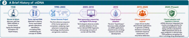
![]() Figure 2. A brief history of ctDNA. The ctDNA field has progressed from the discovery of cell-free DNA in blood to sensitive genomic assays, longitudinal disease monitoring, molecular residual disease assessment, and prospective evaluation in clinical trials.
Figure 2. A brief history of ctDNA. The ctDNA field has progressed from the discovery of cell-free DNA in blood to sensitive genomic assays, longitudinal disease monitoring, molecular residual disease assessment, and prospective evaluation in clinical trials.
A tissue biopsy remains an important source of information about tumor histology and molecular biology. However, a tissue sample represents one location at one point in time. Obtaining another biopsy may also be difficult, particularly when the tumor is inaccessible, available tissue is limited, or repeated assessments are needed.
A blood-based ctDNA measurement offers several complementary advantages:
- Minimally invasive collection: Blood sampling is generally more practical for repeated measurements than serial tissue biopsies.
- Longitudinal assessment: Serial samples can show how ctDNA changes during or after treatment.
- Genomic profiling: ctDNA may identify tumor-associated alterations that can inform targeted treatment selection.
- Resistance monitoring: Newly detected alterations may provide evidence of tumor evolution or an emerging resistance mechanism.
- Assessment across disease sites: DNA released from different tumor deposits may contribute to the circulating signal, potentially providing information beyond a single sampled lesion.
1. Diagnosis and molecular profiling
Although ctDNA is not recommended as a stand-alone diagnostic test for most cancers, it can complement tissue testing when tumor material is limited or difficult to obtain. In advanced solid tumors such as non-small cell lung cancer, plasma-based assays are often used to support molecular characterization; however, negative results may require tissue confirmation because not all tumors shed detectable ctDNA.
2. Treatment selection and genomic profiling
One of the most established uses of ctDNA is the identification of actionable tumor alterations. When sufficient tumor-derived DNA is present, a plasma assay can provide molecular information that may support treatment selection or clinical-trial enrollment.
ctDNA testing may also help identify molecular changes that arise after treatment and provide information about possible resistance mechanisms.
3. Monitoring treatment response
Because ctDNA can be measured repeatedly, investigators can assess whether the circulating tumor signal decreases, clears, persists, or increases during treatment.
A decrease may indicate reduced tumor-derived material in circulation, whereas persistence or an increase may be associated with residual or progressing disease. Importantly, the interpretation depends on the assay, the tumor type, the treatment, the timing of collection, and the specific ctDNA metric being evaluated. Serial measurements are therefore generally more informative than treating ctDNA as a simple one-time positive-or-negative result.
4. Molecular residual disease
After surgery or another curative-intent treatment, imaging may show no detectable disease even though a small number of cancer cells remain. The use of ctDNA to identify this molecular signal is commonly discussed in the context of
molecular residual disease, or MRD.
The potential clinical value is substantial: ctDNA could help distinguish patients who remain at higher risk of recurrence from patients who may already have received sufficient treatment. However, decisions to escalate or de-escalate therapy require careful validation because false-positive and false-negative results can have meaningful consequences.
The FDA's November 2024 final guidance [4] addresses ctDNA use in early-stage, curative-intent solid-tumor drug development, including patient selection or stratification, MRD assessment, response monitoring, and evaluation as an early clinical-trial endpoint. The guidance does not address cancer screening, diagnosis, or the metastatic setting.
5. Recurrence and resistance surveillance
Serial ctDNA testing after treatment may provide molecular evidence of recurrence before disease is clinically apparent through conventional methods. In this setting, a newly detectable or rising ctDNA signal can indicate that closer follow-up or confirmatory evaluation may be warranted. The appearance of new tumor-derived alterations may also reveal emerging resistance before it is fully reflected by imaging or symptoms.

![]() Figure 3. Where ctDNA fits in the patient journey. Potential applications span the cancer-care continuum, from genomic profiling and treatment selection to on-treatment monitoring, molecular residual disease assessment, and recurrence or resistance surveillance.
Figure 3. Where ctDNA fits in the patient journey. Potential applications span the cancer-care continuum, from genomic profiling and treatment selection to on-treatment monitoring, molecular residual disease assessment, and recurrence or resistance surveillance.
Although ctDNA has already demonstrated substantial clinical utility, it is still in the early stages of realizing its full potential. The next generation of ctDNA technologies is focused on improving assay sensitivity, expanding the biological information that can be extracted from a blood sample, and establishing ctDNA as a reliable biomarker for clinical decision-making and drug development.
One of the greatest challenges in ctDNA analysis is that tumor-derived DNA often represents only a tiny fraction of the total cell-free DNA present in circulation. This is particularly problematic in early-stage disease, after surgical resection, or during periods of deep response to therapy, when only a few tumor-derived molecules may be present in a standard blood sample. Consequently, ongoing technological innovation is largely centered on improving the ability to detect increasingly rare ctDNA signals while minimizing background noise from normal DNA and technical artifacts.
Tumor-informed vs. tumor-agnostic assays
One established way to address assay sensitivity is to distinguish between tumor-informed and tumor-agnostic approaches.
Tumor-informed assays begin with sequencing a patient's tumor tissue to identify a personalized set of mutations. Those specific mutations are then tracked in plasma over time. Because the assay is designed around known tumor-specific alterations, this approach can achieve very high sensitivity.
In contrast,
tumor-agnostic (or tumor-naïve) assays do not require a tissue specimen. Instead, they search directly for cancer-associated genomic signals within plasma. These approaches are operationally simpler and may be more scalable across clinical settings, but historically have faced greater sensitivity challenges when disease burden is extremely low.
DNA methylation
Historically, most ctDNA assays have focused on detecting tumor-associated mutations. However, cancer alters far more than DNA sequence alone.
One of the most rapidly evolving areas of research involves
DNA methylation, an epigenetic modification that regulates gene expression and can exhibit highly tumor-specific patterns. Because methylation changes often occur early in tumor development and can be present across large regions of the genome, methylation-based approaches may provide greater sensitivity than mutation-based approaches alone.
Importantly, methylation profiles may also provide information about a tumor's tissue of origin, opening possibilities for identifying where a cancer signal originated rather than simply detecting its presence.
ctDNA offers a dynamic measurement of tumor-associated biology that could help strengthen the connection of dose, drug exposure, biological response, and efficacy. Potential clinical-pharmacology applications include:
- Relating exposure to longitudinal ctDNA changes
- Comparing ctDNA dynamics across dose levels or regimens
- Exploring heterogeneity in response
- Supporting patient stratification
- Complementing imaging and conventional clinical endpoints
- Informing early development decisions when mature survival data are unavailable
- Contributing to dose-optimization and model-informed drug-development strategies
One especially relevant example from the ASCPT journal family is the 2022 Clinical and Translational Science article by Janssen and colleagues [5]. The investigators used longitudinal nonlinear mixed-effects modeling to characterize EGFR-mutant ctDNA measurements from patients with non-small-cell lung cancer and connected the longitudinal model with a parametric progression-free-survival model. The analysis illustrates how pharmacometric methods can move beyond a single ctDNA measurement and quantitatively characterize biomarker dynamics over time.
- Wan JCM, Massie C, Garcia-Corbacho J, et al. Liquid biopsies come of age: towards implementation of circulating tumour DNA. Nature Reviews Cancer. 2017.
- Heitzer E, Haque IS, Roberts CES, Speicher MR. Current and future perspectives of liquid biopsies in genomics-driven oncology. Nature Reviews Genetics. 2019.
- Ignatiadis M, Sledge GW, Jeffrey SS. Liquid biopsy enters the clinic: implementation issues and future challenges. Nature Reviews Clinical Oncology. 2021.
- U.S. Food and Drug Administration. Use of Circulating Tumor Deoxyribonucleic Acid (ctDNA) for Early-Stage Solid Tumor Drug Development: Guidance for Industry. Silver Spring, MD: U.S. Food and Drug Administration; November 2024.
- Janssen JM, Verheijen RB, van Duijl TT, et al. Longitudinal nonlinear mixed effects modeling of EGFR mutations in ctDNA as predictor of disease progression in treatment of EGFR-mutant non-small cell lung cancer. Clinical and Translational Science. 2022.
